Educational Chart
Paramoecium Chart
Laminated 58 × 90 cm wall chart showing structure, nutrition, locomotion and reproduction — ideal for the classroom wall.
₹170 (Exc. GST)
View product

+91 8683 878 878
Pan India Delivery


Paramecium (also spelt Paramoecium) is a microscopic, single-celled organism that lives in freshwater. It belongs to Kingdom Protista and the phylum Ciliophora, and its slipper-shaped body is covered in thousands of tiny hair-like cilia that it uses to swim and to sweep in food. It is one of the most studied organisms in biology, which is why a labelled Paramecium diagram appears in almost every school exam.
Paramecium is a tiny, free-living organism made of just one cell. You will not see it with the naked eye — a drop of pond water under a school microscope is usually where students meet it first, moving quickly across the slide. Despite being a single cell, it behaves like a complete little animal: it swims, hunts for food, digests it, balances its water content and reproduces, all on its own.
It is grouped under Kingdom Protista, the kingdom set aside for single-celled eukaryotes that do not fit neatly into plants, animals or fungi. Within Protista it belongs to the phylum Ciliophora — the ciliates — named after the thousands of cilia that cover the cell. Paramecium lives mostly in stagnant freshwater such as ponds, ditches and puddles, where it feeds on bacteria and decaying matter.
Biologists study Paramecium so often that it has been called the “white rat” of its phylum. It is easy to grow, easy to see and behaves in predictable ways — which is also exactly why it shows up year after year in school and competitive biology exams.
There is no difference: both words name the same organism. ‘Paramecium’ is the modern, internationally accepted spelling, while ‘Paramoecium’ (from the older Latinised form Paramœcium) is still printed in many Indian textbooks and asked in exams. If your question paper says “Paramoecium,” it is asking about the very same slipper-shaped ciliate described here.
The labelled diagram above shows the main parts of a Paramecium. The table below explains what each part does — a quick way to revise for the “label the diagram” and “state the function” questions.
| Part | Function |
|---|---|
| Cilia | Thousands of short hair-like projections covering the body; beat in rhythm for locomotion and to draw food in. |
| Pellicle | The flexible but firm outer covering that holds the cell’s shape while allowing movement. |
| Oral groove | A slanting channel on one side, lined with cilia, that sweeps food particles toward the cell mouth. |
| Cytostome (cell mouth) | The opening at the base of the oral groove through which food enters the cell. |
| Cytopharynx (gullet) | A short funnel beyond the cytostome where food collects before a food vacuole forms. |
| Food vacuole | A bubble of food and water pinched off from the gullet; digestion happens inside it as it circulates. |
| Contractile vacuoles | Usually two star-shaped vacuoles that collect excess water and pump it out (osmoregulation). |
| Macronucleus | The large kidney-shaped nucleus controlling metabolism, growth and everyday cell activity. |
| Micronucleus | The small nucleus that stores genetic material for reproduction and conjugation. |
| Trichocysts | Tiny defensive bodies under the pellicle that can fire out thread-like filaments. |
| Cytoproct (anal pore) | A fixed spot where undigested waste is expelled from the cell. |
The whole body is wrapped in a thin, firm layer called the pellicle, which keeps the slipper shape while still bending as the cell moves. Embedded just beneath it are the cilia — several thousand short, hair-like threads arranged in neat rows over the entire surface.
On one side runs the oral groove, a slanting, ciliated channel that leads to the cytostome (the cell mouth). Beyond the mouth is the cytopharynx, or gullet, where incoming food gathers. The clear outer layer of cytoplasm is the ectoplasm and the inner, granular layer is the endoplasm, which holds the cell’s organelles.
Floating in the cytoplasm are food vacuoles (where digestion happens) and usually two contractile vacuoles, one near each end, that look like tiny stars because of their radiating canals. Because Paramecium lives in freshwater, water constantly seeps in; the contractile vacuoles collect this surplus and squeeze it back out, keeping the cell from bursting. Two nuclei sit near the centre: the large, kidney-shaped macronucleus and the small micronucleus tucked beside it. Scattered under the pellicle are trichocysts, defensive bodies that can shoot out fine threads when the cell is threatened.
Here is where Paramecium sits in the modern biological classification:
| Rank | Group |
|---|---|
| Domain | Eukaryota |
| Kingdom | Protista |
| Phylum | Ciliophora (ciliates) |
| Class | Oligohymenophorea |
| Order | Peniculida |
| Family | Parameciidae |
| Genus | Paramecium (O. F. Müller, 1773) |
| Common species | P. caudatum, P. aurelia, P. bursaria |
Paramecium swims using its cilia. The cilia do not all beat at once — they move in smooth, travelling waves, rather like rows of oars dipping one after another. This pushes the cell forward in a gentle spiral while it spins slowly on its own axis. If it runs into an obstacle or an unpleasant chemical, it performs an avoiding reaction: it briefly reverses the beat of its cilia, backs up, turns slightly and tries a new direction. This simple trial-and-error steering lets a single cell navigate its world surprisingly well.
Paramecium is a heterotroph — it cannot make its own food, so it eats other microbes such as bacteria, yeasts and algae. The cilia lining the oral groove beat to create a current that sweeps food particles, along with a little water, down into the cytostome. From there the food passes into the gullet, where it is packed into a food vacuole. The vacuole then circulates through the cytoplasm (a movement called cyclosis) while digestive enzymes break the food down and the nutrients are absorbed. Whatever cannot be digested is carried to the cytoproct, a fixed anal pore, and pushed out of the cell.
Paramecium reproduces in more than one way:
Because it is large for a single cell, lives in ordinary pond water and moves visibly under low magnification, Paramecium is one of the first organisms a biology student observes for themselves. Seeing the real thing swim — and then matching it to a labelled chart or model — is what makes the structure stick. The right teaching aids turn an abstract diagram into something a class can actually point to and discuss.
Laminated 58 × 90 cm wall chart showing structure, nutrition, locomotion and reproduction — ideal for the classroom wall.
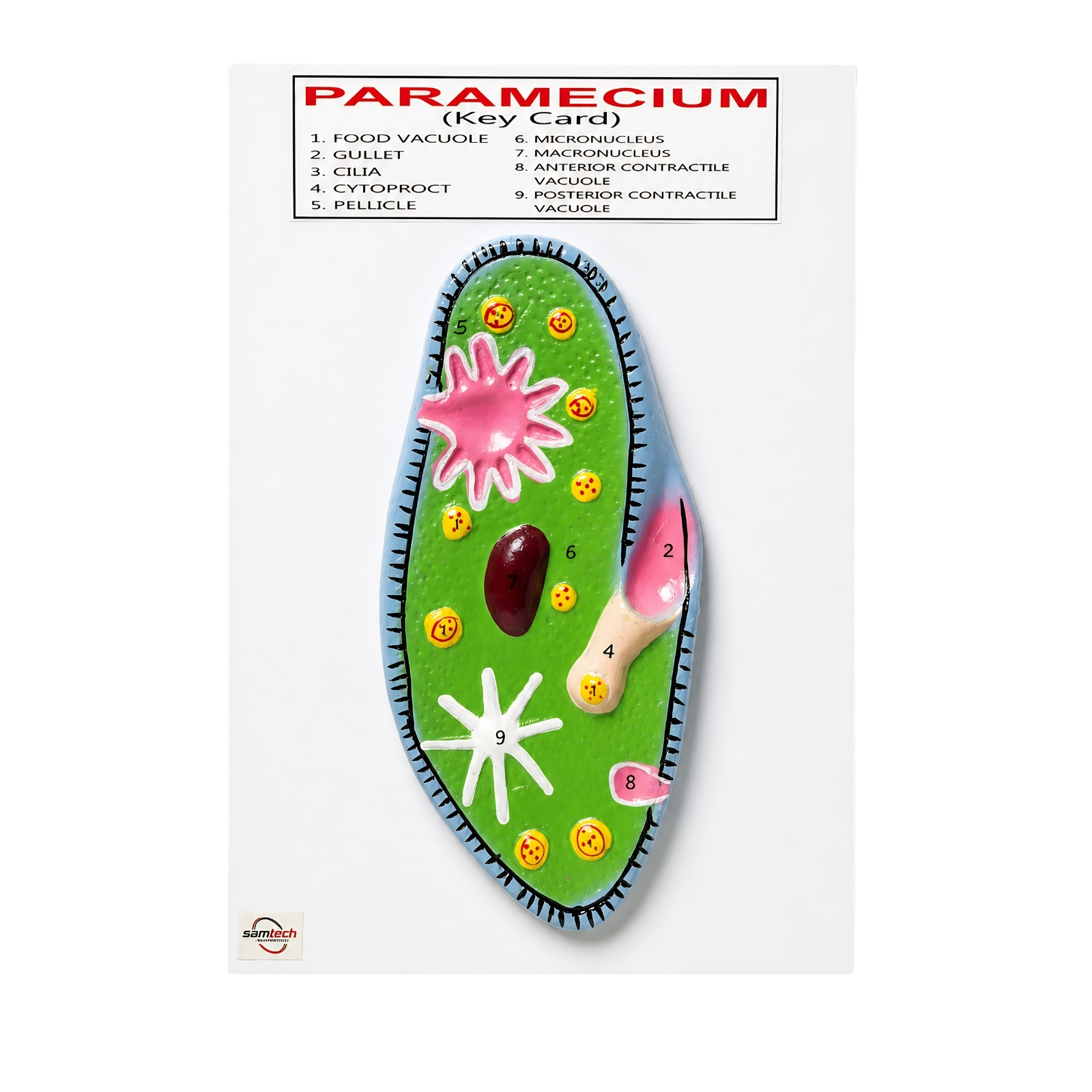
Enlarged, colour-coded 3D model with 9 clearly labelled parts — turns the diagram into something a class can hold and point to.
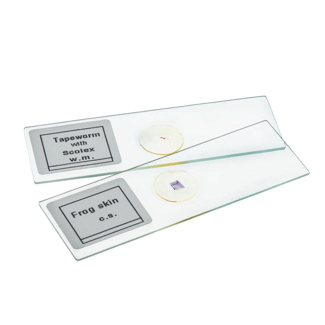
Prepared slide set for the biology lab — Paramecium is one of the specimens, ready to view live under the microscope.
Schools, dealers and GeM buyers — these items are manufactured in India and supplied in bulk (MOQ 50). For dealer pricing or a quotation, call +91 8683 878 878.
Neither. Paramecium is a protist — it belongs to Kingdom Protista, a separate group for single-celled eukaryotes that are not plants, animals or fungi. It feeds like an animal (it eats other microbes) but is far simpler than any animal.
Because its single cell is shaped like the sole of a slipper or shoe — broad and rounded at the front and slightly tapered at the back. ‘Animalcule’ is an old word for a tiny living thing seen under a microscope.
The large macronucleus controls the cell’s everyday activities such as growth, feeding and metabolism. The small micronucleus stores the genetic material used during reproduction and conjugation. A Paramecium can survive short-term without the micronucleus but not without the macronucleus.
It swims using its cilia, which beat together in coordinated waves like rows of tiny oars. This drives the cell forward in a spiral path while it rotates on its own axis. When it bumps into an obstacle it briefly reverses the beat of its cilia and changes direction.
It is a heterotroph that eats bacteria, algae and other tiny particles. Cilia in the oral groove sweep food and water into the cytostome (cell mouth), then into the gullet, where a food vacuole forms. Enzymes digest the food inside the vacuole, and undigested waste leaves through the cytoproct.
Paramecium is strictly unicellular — the entire organism is one cell. All its functions (movement, digestion, reproduction, water balance) are handled by specialised structures inside that single cell.
For more detail on the biology of Paramecium, these reference sources are a good next step:

A clear, labelled moving-coil galvanometer diagram with its working principle, construction, formula, types, sensitivity and laboratory uses — exam-ready notes for students.

Ask any science teacher what separates a lesson that sticks from one that doesn’t, and the answer is almost always

Science doesn’t happen in textbooks. It happens when a student holds a Vernier caliper for the first time and actually
No account yet?
Create an Account