







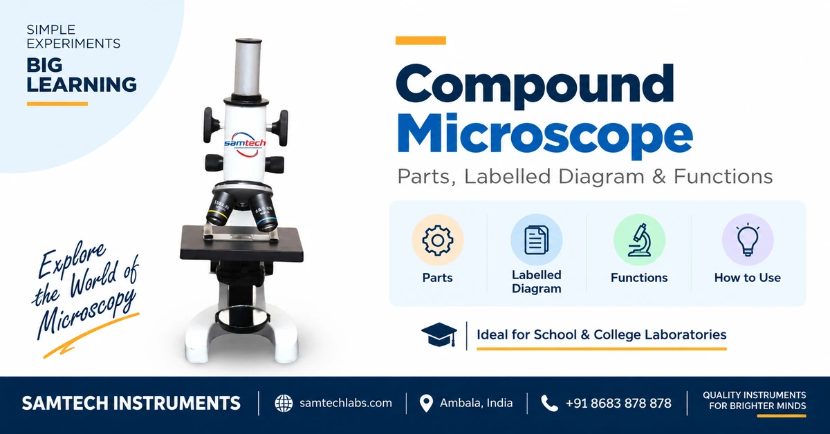

A compound microscope is a laboratory instrument that uses two sets of lenses — an eyepiece and objective lenses — to magnify tiny, thin, transparent specimens such as cells, bacteria and tissues by up to 1000 times. It is the standard high-power microscope used in school and college biology labs. Samtech Instruments has manufactured laboratory microscopes and optical instruments in Ambala, India since 2002, supplying schools, colleges and institutions across India and 30+ countries.
This guide covers everything a student, teacher or lab in-charge needs: the parts of a compound microscope and their functions, a clear labelled diagram, how magnification works, a step-by-step how-to-use method, the difference between compound and other laboratory microscopes, and a short buying guide.
Key takeaways
- A compound microscope uses two lenses in series (eyepiece + objective) — that is why it is called “compound”.
- Total magnification = eyepiece × objective. A 10× eyepiece with a 100× objective gives 1000×.
- It has optical parts (lenses, condenser, diaphragm, illuminator) and mechanical parts (arm, stage, knobs, base).
- Use the coarse knob to focus at low power and only the fine knob at high power.
- It views thin, transparent specimens on slides — a dissecting microscope is used for whole, solid objects.
📑 Table of contents
What is a compound microscope?
A compound microscope is a high-power optical microscope that magnifies an object in two stages. The objective lens (near the specimen) forms a magnified real image, and the eyepiece lens (near your eye) magnifies that image again. Because two lens systems act one after the other, the magnifying powers multiply — letting you see structures as small as bacteria.
It works only on specimens that are thin enough for light to pass through, mounted on a glass microscope slide and protected by a cover glass. This is why it is the workhorse of biology labs for viewing cells, blood, pond water organisms like Paramecium, and prepared tissue slides.
Labelled diagram of a compound microscope
Parts of a compound microscope and their functions
A standard compound microscope has around 14 key parts. The table below lists each one with its function — the format most useful for revision and lab practicals.
| Part | Function |
|---|---|
| Eyepiece (ocular lens) | The lens you look through, usually 10×. It magnifies the image formed by the objective. |
| Body tube (head) | Connects the eyepiece to the objectives and carries the light path between them. |
| Arm | The curved support that holds the tube and stage; you hold the arm when carrying the microscope. |
| Revolving nosepiece | Rotating turret holding the objectives; click a new objective into place to change magnification. |
| Objective lenses | Lenses closest to the specimen (4×, 10×, 40×, 100×) that do the primary magnification. |
| Stage | Flat platform with a central hole where the slide is placed for viewing. |
| Stage clips | Spring clips that hold the slide firmly in position on the stage. |
| Diaphragm (iris) | Adjustable opening below the stage that controls how much light reaches the specimen. |
| Condenser | Lens that gathers and focuses light from the illuminator onto the specimen. |
| Coarse adjustment knob | Large knob that moves the stage/tube quickly for rough focusing at low power. |
| Fine adjustment knob | Small knob for tiny movements that give a sharp final image; the only knob used at high power. |
| Illuminator / mirror | Built-in LED light (or a mirror) that supplies light to view the specimen. |
| Nose / draw tube | Holds the eyepiece and sets the correct optical tube length. |
| Base (foot) | Heavy bottom that gives the microscope stability on the bench. |
Optical parts vs mechanical parts
Examiners often ask students to classify the parts. Here is the simple split:
| Optical parts (make & light the image) | Mechanical parts (support, hold & focus) |
|---|---|
| Eyepiece, objective lenses, condenser, diaphragm, illuminator/mirror | Base, arm, stage, stage clips, body tube, nosepiece, coarse & fine adjustment knobs |
How magnification works
Total magnification is simply the eyepiece power multiplied by the objective power:
| Eyepiece | Objective | Total magnification | Typical use |
|---|---|---|---|
| 10× | 4× (scanning) | 40× | Finding the specimen, whole-mount view |
| 10× | 10× (low power) | 100× | Tissue sections, plant/animal cells |
| 10× | 40× (high power) | 400× | Cell detail, nucleus, blood cells |
| 10× | 100× (oil immersion) | 1000× | Bacteria, fine detail (with immersion oil) |
How to use a compound microscope (step by step)
- Place and power onSet it on a flat, steady bench. Switch on the LED illuminator (or angle the mirror) to a low brightness.
- Start on the lowest objectiveRotate the nosepiece so the 4× or 10× objective clicks into place.
- Mount the slidePut the prepared slide on the stage, specimen over the light hole, and secure it with the stage clips.
- Focus with the coarse knobLooking through the eyepiece, turn the coarse adjustment slowly until the specimen appears.
- Set the lightOpen or close the diaphragm for even lighting and good contrast.
- Sharpen with the fine knobUse only the fine adjustment to make the image crisp.
- Go to higher powerSwitch to 40× for detail and re-focus with the fine knob only — never the coarse knob at high power.
- Shut down & cleanReturn to the lowest objective, lower the stage, wipe lenses with lens tissue and cover the microscope.
Compound microscope vs other laboratory microscopes
Schools and colleges usually keep more than one kind of microscope. Here is how the compound microscope compares with the others Samtech supplies:
| Microscope | Magnification | Best for | Image |
|---|---|---|---|
| Compound | 40×–1000× | Thin, transparent specimens on slides (cells, bacteria, tissue) | Flat 2D, inverted |
| Dissecting (stereo) | 10×–40× | Whole, solid, opaque objects; dissection, insects, coins | Upright 3D |
| Travelling | Low, with vernier scale | Physics measurement of small distances & refractive index | Measuring, not viewing |
| Projection | Medium | Showing one image to a whole class on a screen | Projected |
| Digital / trinocular | 40×–1000× | Photography, recording & display via camera | On monitor |
Which microscope should you buy?
🏫 Schools (Class 6–12)
A monocular/binocular student compound microscope with 4×, 10×, 40× objectives and LED light covers the full biology syllabus affordably.
Student Compound Microscope →🧪 Colleges & labs
Go for a research-grade binocular compound with 100× oil-immersion for bacteriology and advanced practicals.
Compound Microscope →🔎 Dissection & specimens
For whole insects, flowers and dissection work, add a low-power stereo dissecting microscope.
Dissecting Microscope →💻 Demonstration & digital
Need to show the whole class or record images? Choose a projection or USB-camera trinocular model.
Digital Trinocular →Don’t forget consumables and accessories: glass slides, cover slips, prepared slides and a spare LED lamp.
Care & maintenance
Well-maintained optics last for decades. Carry the microscope with one hand on the arm and one under the base; clean lenses only with lens tissue (never cloth or tissue paper); remove immersion oil immediately after use; store it covered on the lowest objective; and keep it in a dry, dust-free cabinet with silica gel during humid months.
Buying microscopes for your school or college?
Samtech Instruments manufactures compound, student, dissecting, travelling and digital microscopes in Ambala — with bulk pricing for institutions and full after-sales support.
Why choose Samtech Instruments
A physics and biology instrument manufacturer based in Ambala, India — the country’s hub for laboratory equipment — supplying institutions since 2002.